Wednesday, December 27, 2006

Lax Editorial Standards At Nature Neuroscience?

You be the judge...
Editorial
Nature Neuroscience - 10, 1 (2007)
Setting the record straight

The discovery of serious errors in two recent papers in the journal leads to lessons for authors, referees and editors.

. . .

The first correction involves a Brief Communication (Makara et al., 2005) reporting that inhibition of the enzyme that breaks down the endocannabinoid 2-arachidonoylglycerol enhances retrograde signaling in the hippocampus. The authors concluded that 2-arachidonoylglycerol is important for synaptic plasticity and that the enzyme is a possible drug target, in part because one of the putative inhibitors tested appeared to be specific for the enzyme. They subsequently discovered that
the commercial preparation of this drug was contaminated. When the contaminant was eliminated, the effect disappeared. . . .

The second correction is more complex. The original article (Grill-Spector et al., 2006) reported high-resolution fMRI measurements in the fusiform face area (FFA), a region of the visual cortex that responds more to faces than to other visual stimuli. The authors drew two conclusions: that the FFA is heterogeneous, in that the degree of selectivity varies over the region, and—more remarkably—that the FFA contains some voxels that are highly selective for object categories other than faces. After the paper was published, two groups wrote to point out flaws in the analysis. One letter (Simmons et al., 2007) noted that
the authors used a formula for selectivity that erroneously assigns high selectivity values to voxels with negative responses to nonpreferred categories, causing a substantial overestimate in selectivity for all object categories.

Another group (Baker et al., 2007) spotted a more subtle flaw:
the analysis used to demonstrate selectivity for particular categories did not distinguish between random variation and replicable effects reflecting neural tuning. Random variation can cause some voxels to respond more to some categories than to others. To demonstrate that such differences reflect neural selectivity requires an appropriate statistical analysis, for instance cross-validation across independent datasets. The original paper seemed to report the results of such an analysis—that voxel selectivity was highly correlated between even and odd scans. However, communication with the authors revealed that this analysis had excluded voxels whose responses were negatively correlated across the two sets of scans, a detail that was omitted from the paper. This restriction could falsely increase consistency across scans. Indeed, when the authors redid their analysis without it, the selectivity for nonface objects was not replicated from one set of scans to the next.
My focus is on the second paper, because (1) I can't say anything about 2-arachidonoylglycerol, and (2) getting a contaminated drug from a vendor is not the authors' fault.

The editorial continues:
The authors of the article acknowledge both errors in their correction (Grill-Spector et al., 2007). When these errors are fixed, the most interesting conclusion of the paper—that the FFA contains voxels highly selective for nonface objects—is no longer supported.
However, the editors refuse to retract the paper:
In both cases, after considerable discussion with colleagues, we have decided to publish a correction to the original paper rather than a retraction, even though it seems likely that neither paper would have been published in Nature Neuroscience had the errors been identified and corrected during the review process. Retractions were deemed inappropriate because they would have removed from the record some valid data and conclusions that are likely to be useful to specialists in the field, and it seemed unlikely that the authors would be able to publish these data elsewhere.
Well, boo hoo!! The authors can't publish their compromised data anywhere except in Nature Neuroscience. Let's all send our unpublishable data to Nature Neuroscience! We can also appeal to the archenemy journal, Science, to publish results that contradict articles in the Nature family of journals...

The Neurocritic started this blog as a means of quality control in the field, a way to point out errors, exaggerations, misinterpretations of data, or as the masthead says,

Deconstructing the most sensationalistic recent findings in Human Brain Imaging, Cognitive Neuroscience, and Psychopharmacology

The Nature Neuroscience editorial concludes:
...the ultimate responsibility for recruiting referees with appropriate expertise lies with the editors, and in this case we clearly should have consulted referees with stronger mathematical expertise.

Nonetheless, it is common practice in functional imaging (and indeed in other areas of neuroscience) to analyze experiments by
selecting data according to some criteria and then plotting the average response, without testing an independent data set to ensure that the selection criteria have not merely picked out random variation in a particular direction.
How many other prominent articles are of dubious accuracy? The saga continues...

Subscribe to Post Comments [Atom]

Sunday, December 24, 2006

Seasonal, Inspiring, Humorous, Entertaining, and Bizarre

In case it wasn't clear from the last two posts:
US Highlights
Douglas Kamerow, US editor

As usual, the Christmas double issue of the BMJ is seasonal, inspiring, humorous, entertaining, and bizarre. There is too much good stuff to describe it all, but here are some highlights.
Additional highlights for me include:
You're not going to give me the umbrella, are you? C Bradbeer, S Soni, A Ekbote, T Martin. BMJ 2006;333:1287-1288.

The "umbrella test" is a longstanding urban myth that still bothers men who present for testing at sexual health clinics.
AND
The ideal tool for decorators: a novel use for disposable laryngoscope blades. RJ Green, JMT Pierce. BMJ 2006;333:1297-1298.

We noticed that the tip of a Timesco (London, UK) Callisto Mackintosh disposable laryngoscope blade fitted perfectly between the rim and lid of a tin of paint. This, coupled with the smooth contour, which fitted comfortably into the hand, led us to investigate which of the three commercially available blade sizes (2, 3, and 4) needed the least force to open tin lids. The null hypothesis for the study was that the forces needed would not differ.


View the entire table of contents!

Subscribe to Post Comments [Atom]

Saturday, December 23, 2006

Sword Swallowing And Its Side Effects


Just when you thought it couldn't get any stranger in the latest issue of the British Medical National Enquirer, er, Journal:
Sword swallowers more likely to be injured when distracted or swallowing "unusual" swords

Sword swallowers are more likely to sustain an injury--such as a perforation of the oesophagus-- if they are distracted or are using multiple or unusual swords [
NO! Really??], finds a study in this week's Christmas issue of the BMJ.

Sword swallowers who can swallow a non-retractable solid steel blade at least two centimetres wide and 38 centimetres long are recognised by the SSAI (Sword Swallowers' Association International). Despite the obvious dangers of the profession, English medical literature contains only two case reports of injury resulting from sword swallowing.
Other words of wisdom from the original article by Witcombe & Meyer:
Sword swallowers know their occupation is dangerous.

Sore throats are common, particularly while the skill is being learnt or when performances are too frequent.

Sword swallowers without healthcare coverage expose themselves to financial as well as physical risk.
Oh, but it gets even better!!


One of the authors (DM) swallowing seven swords

Read more!

Witcombe B, Meyer, D (2006). Sword swallowing and its side effects. BMJ 333: 1285-1287.

To the Hilt Sword Swallowing

Subscribe to Post Comments [Atom]

Are Surgeons Taller And Better Looking Than Other Doctors?


Is your family practice doc ugly? Is your surgeon hot? Is George Clooney better looking than both of them? Now you know why!!
Surgeons are taller and more handsome than physicians, finds a study in this week's Christmas issue of the BMJ.

Doctors at the University of Barcelona Hospital noticed that the tallest and most handsome male students were more likely to go for surgery, and the shortest (and perhaps not so good looking) ones were more likely to become physicians.
Read more!

Trilla A, Aymerich M, Lacy AM, Bertran MJ (2006). Phenotypic differences between male physicians, surgeons and film stars: comparative study. BMJ 333:1291-1293.
Conclusions Male surgeons are taller and better looking than physicians, but film stars who play doctors on screen are better looking than both these groups of doctors. Whether these phenotypic differences are genetic or environmental is unclear.
Here's the Introduction in its entirety:
We finished our medical training at the University of Barcelona more than 25 years ago, and have enjoyed our work ever since. At medical school we noted certain differences between male trainees who selected either surgery or medicine as their specialty. The tallest and most handsome male students were more likely to go for surgery, and the shortest (and perhaps not so good looking) ones were more likely to become physicians (including doctors of internal medicine and its subspecialties).

Now, after all these years we hypothesise that, on average, surgeons are taller and better looking than physicians. We conducted a comparative study to test this hypothesis.
From the Results:
We did not make individual results public. However, widespread rumours, discussions, polls, and illegal bets arose throughout the institution as a by-product of our study. If they requested, participants were privately told about their personal score compared with the average score of the relevant group.

Sample of participants. Left, surgeon; middle, physician; right, control (George Clooney)

And in the Acknowledgments section:
Competing interests: AT is a physician and AML is a surgeon. AT and MA have been happily married for 25 years. MA's good looking score for AT was not requested to avoid any problems at home for Christmas.
The real question is, will Pedro Almodovar adapt this paper for the big screen?? It might be a bit of a stretch for him, since most of his recent films are about women...

Subscribe to Post Comments [Atom]

Friday, December 22, 2006

Wine and the Brain -- Dehumanizing Wine

An interesting thread at Mark Squires' Bulletin Board on eRobertParker.com, started by Edward Comstock, Oenophile:

Wine and the Brain--dehumanizing wine
At the risk of seeming argumentative, I would rather like to belabor a point raised in a recent thread about the neuroscience of pleasure (as it involves wine drinking), as it is painful to me to see the humanity vacuumed out of such a human enterprise as wine drinking, through the common arguments of the acolytes of bio-reductivism.
These reductive theories of the brain’s need to pursue pleasure and avoid pain suppose that the purpose and essence of the mind is to regulate and represent the sensations and the physical state of the body, such that ideas and mental activity is directed at this physical state. Therefore, it is claimed, my brain is “hard-wired” to want to find 1990 La Tache or the like because it brings me an emotional state which is a response to the physical sensations--the chemical-based impressions--provided by the La Tache. My emotion of “ecstasy” in drinking the La Tache is the same thing as the physical (or neurological) state, a release of dopamine for example, provided from drinking the La Tache, it is claimed.

This idea is used to undermine the idea that critics such as Parker do not describe properties of the wine, but rather personal sensations; and this idea is false. Taste, before it is a physical impression, is a thing in the world--and tasting is a performance, super-charged with ritual and cultural modalities.
And this press release, based on a new paper in Neuron:
How Your Brain Helps You Become A Wine Expert

You don't need to sign up for pricey wine appreciation classes to parse the subtle difference between the black cherry bouquet of a pinot noir and the black currant scent of a cabernet sauvignon. Just pour yourself a couple glasses and sniff. Your brain will quickly help you become a modest oenophile. It's up to you if you want to drink the lesson plan.

A new study by Northwestern University researchers shows that the brain learns to differentiate between similar smells simply through passive experience, shedding light on how we ultimately learn to identify thousands of smells from birth. The study also revealed for the first time how and where the brain modifies and updates information about smells.
Reference

Li W, Luxenberg E, Parrish T, Gottfried JA. (2006). Learning to smell the roses: experience-dependent neural plasticity in human piriform and orbitofrontal cortices. Neuron 52:1097-108.

Subscribe to Post Comments [Atom]

Thursday, December 21, 2006

Positive Voltage Does Not Equal Excitation. Or Worse, Positive Emotion!

Oh no!
Reading Shakespeare Has Dramatic Effect On Human Brain

Research at the University of Liverpool has found that Shakespearean language excites positive brain activity, adding further drama to the bard's plays and poetry.

Shakespeare uses a linguistic technique known as functional shift that involves, for example using a noun to serve as a verb. Researchers found that this technique allows the brain to understand what a word means before it understands the function of the word within a sentence. This process causes a sudden peak in brain activity and forces the brain to work backwards in order to fully understand what Shakespeare is trying to say.

. . .

Professor Neil Roberts, from the University’s Magnetic Resonance and Image Analysis Research Centre, (MARIARC), explains: “The effect on the brain is a bit like a magic trick; we know what the trick means but not how it happened. Instead of being confused by this in a negative sense, the brain is positively excited. The brain signature is relatively uneventful when we understand the meaning of a word but when the word changes the grammar of the whole sentence, brain readings suddenly peak. The brain is then forced to retrace its thinking process in order to understand what it is supposed to make of this unusual word.”
This study was covered in a similar manner in an APA-sponsored site! How embarrassing for them!
Reading Shakespeare excites the brain
United Press International - December 19, 2006

LIVERPOOL, England, Dec 19, 2006 (UPI via COMTEX) -- British researchers have found reading Shakespeare has a dramatic effect on the human brain, producing positive brain activity.

To explain a little, the researchers recorded EEG while participants read selections from Shakespeare. They were looking for EEG signatures of semantic violations (indexed by a negative voltage brain wave at ~400 msec, called the N400) and syntactic violations (indexed by a positive-voltage brain wave at ~600 msec, called the P600).
Above figure from a different study, published in Biological Psychology by Isel et al. (2006)

The brain waves were obtained by averaging a bunch of EEG trials together, and these event-related potential (ERP) components reflect summed electrical activity (post-synaptic potentials) from a huge number of pyramidal cells, recorded remotely from the scalp (to put it simply). The polarity of these components (i.e., positive or negative) does not indicate whether they are excitatory or inhibitory.

Where were these Shakespeare ERP results published?? According to the UPI story, the study appeared in The Reader, a literary magazine (although I couldn't find it...).

Subscribe to Post Comments [Atom]

Tuesday, December 19, 2006

Science For Hip Teens

Moving right along with the TV analogies, this LiveScience.com article (below) reminded me of the Simpsons episode where a new character, Poochie the dog, is introduced into The Itchy and Scratchy Show in a misguided effort to appeal to kids and to boost declining ratings.

Teen Talk: Science Needs to Dazzle
By Jeanna Bryner
posted: 19 December 2006

SAN FRANCISCO - Scientists who want to get their messages across to hip teens should spice up their presentations without dumbing down the science.

When it comes to climate change, educating the next generation of earth-saving scientists takes savvy, scientists said here last week at a meeting of the American Geophysical Union.

"Teenagers live in an MTV world so most things they are exposed to are slick and well produced," said Katharine Giles, a research fellow at the Center for Polar Observation and Modelling in the United Kingdom. "So anything like a lecture should try and get to the same standard."

The Faraday Lecture of 2006 did just that. With swirling lights, electronic music, videos and lots of audience participation, more than 30,000 attendees got a dose of "Emission Impossible: Can Technology Save the Planet?"


Compare to:

The Itchy & Scratchy Show: "The Beagle Has Landed"

Itchy and Scratchy are driving to a fireworks factory, when they pass a beagle dressed as a surfer dude. Itchy tells Scratchy, "It's our new friend, Poochie."

To show that he's ultra-cool, Poochie uses a rap to introduce himself. Scratchy thinks Poochie is "one outrageous dude", while Itchy opines the beagle is "totally in my face."

Further revealing his character, Poochie then performs a rock guitar solo, before demonstrating how he can skateboard and play basketball at the same time. Before exiting, he uses some more hip language: "Catch you on the flip side, dudemeisters!" He also feigns a high five to Scratchy, before reminding the audience to recycle to the extreme and taking off in Itchy and Scratchy's car (passing the fireworks factory).


Kids just want to see the cartoon violence, right? Explosions and all?? Can a focus group of adult scientists produce videos that will inform and educate teens about climate change (for example) and yet still appeal to them?
"Teenagers that were surveyed thought that celebrities could make more of a difference to mitigating our effect on the climate than engineers and scientists could," Giles said. "This may well actually be true, but I think it raises quite an important issue. There's a misconception about what scientists and engineers actually do."

Subscribe to Post Comments [Atom]

Today's Disorder: Prader Willi Syndrome

According to the Prader-Willi Syndrome Association (UK):
Prader-Willi Syndrome (PWS) was first described in 1956 by Swiss doctors, Prof. A Prader, Dr A Labhart and Dr H Willi, who recognised the condition as having unique and clearly definable features. These features are:

* Hypotonia: weak muscle tone, and floppiness at birth.
* Hypogonadism: immature development of sexual organs and other sexual characteristics.
* Obesity: caused by excessive appetite and overeating (hyperphagia), and a decreased calorific requirement owing to low energy expenditure levels. (Obesity is not normally a feature of those whose food intake is strictly controlled.)
* Central nervous system and endocrine gland dysfunction: causing varying degrees of learning disability, short stature, hyperphagia, somnolence, and poor emotional and social development.
image from Tokyo Medical University
It's a genetic disorder resulting from a deletion on chromosome 15 (15q11-13) in 70% of the cases. Another 25% are from a maternal uniparental disomy of chromosome 15, and the remainder are due to imprinting defects (Miller et al., 2006).

But it's most known for the symptom of insatiable appetite, and was even featured in an episode of CSI:, in which the dead body of
Jerry Gable (30), a short baby-faced man with an enormous potbelly, is found in a dumpster in back of a popular restaurant, covered in food remnants. Not a happy way to spend Thanksgiving. So why is there a smile on Jerry's face? There are no visible injuries or wounds other than some pasty white marking on his right wrist, and chipped and inflamed fingernails. There's also a trace of silver-metallic residue on his cheek. A wrapper belonging to Aunt Jackpot's Pretzel Stand is found in his back pocket. In autopsy, Dr. Robbins empties an enormous amount of food content. The victim's stomach was distended to six liters in volume: normal capacity is one, with a maximum load of about four. Jerry died from ASPHYXIA due to mechanical compression of the lungs. In other words, he ate himself to death.
It turns out this character had Prader Willi and was a contestant in a hot dog eating contest. Anyway, a new neuroimaging study examined brain activity when participants with Prader Willi and controls viewed pictures of food (and animals and tools) in the scanner.
Miller JL, James GA, Goldstone AP, Couch JA, He G, Driscoll DJ, Liu Y. (2006). Enhanced activation of reward-mediating prefrontal regions in response to food stimuli in prader-willi syndrome. J Neurol Neurosurg Psychiatry. 2006 Dec 8; [Epub ahead of print]

BACKGROUND: Individuals with Prader-Willi syndrome (PWS) exhibit severe disturbances of appetite regulation, including delayed meal termination, early return of hunger after a meal, seeking and hoarding food, and eating of non-food substances. Brain pathways involved in control of appetite in humans are thought to include the hypothalamus, frontal cortex (including orbitofrontal, ventromedial prefrontal, dorsolateral prefrontal, and anterior cingulate areas), insula, and limbic and paralimbic areas. We hypothesized that the abnormal appetite in PWS results from aberrant reward processing of food stimuli in these neural pathways. METHODS: We compared functional MRI (fMRI) blood-oxygen level dependent (BOLD) responses while viewing pictures of food in eight adults with PWS and eight normal weight adults after ingestion of an oral glucose load. RESULTS: Subjects with PWS demonstrated significantly greater BOLD activation in the ventromedial prefrontal cortex than controls when viewing food pictures. No significant differences were found in serum insulin, glucose, or triglyceride levels between the groups at the time of the scan. CONCLUSIONS: Individuals with PWS had an increased BOLD response in the ventromedial prefrontal cortex compared to normal weight controls when viewing pictures of food after an oral glucose load. These findings suggest that an increased reward value for food may underlie the excessive hunger in PWS, and support the significance of the frontal cortex in modulating the response to food in humans. Our findings in the extreme appetite phenotype of PWS support the importance of the neural pathways that guide reward-related behavior in modulating the response to food in humans.
The paper is very short, and seems to have minimal analyses, primarily FOOD vs. REST (looking at a fixation point), and the other categories vs. REST. They didn't correct for multiple comparisons because
...the Bonferonni corrections performed by BrainVoyager 2000 tend to be too conservative for functional neuroimaging analyses.

from Miller et al. (2006)

At any rate, the figure above shows greater activation of visual cortex in controls, and greater activation in the ventromedial prefrontal cortex (VMPFC) in the PWS participants. However, the PWS group also showed substantial activity in the PFC (more dorsally) for the TOOLS vs. REST comparison, unlike the controls. So it's not exactly appropriate to conclude that
increased reward value for food may underlie the excessive hunger in PWS...
since the TOOL stimulus set included both nonfood tools (e.g. hammers, hairdryers) and food-related tools (e.g. spoons, cups) . The authors' suggestion that the ANIMALS vs. REST comparison served as a check against any tool-related reward activity raises the issue of, um, whether chickens and pigs and cows appeared as stimuli.

Read more about PWS.

Subscribe to Post Comments [Atom]

I'm Not In Touch With My Feelings


Today's Weird Mental Disorder: I Feel Nothing. No, Really.
Posted by Randy Dotinga

I just stumbled across a blog called The Neurocritic, run by a 26-ish guy who declares (on the main page, even) that he "descended into a spiral of drug abuse and prostitution" as a teenager.

Huh.

Anyway, he has a post today on brain-scan research into something called alexithimia, which has nothing to do with anyone named Alex.

. . .

Women might think they've seen this phenomenon before -- in every man they've ever met. I'd be offended by that stereotype, except I'm not in touch with my feelings.
Cool. Really.

Subscribe to Post Comments [Atom]

And the First of the Encephala (version 2.0)


The new and improved Encephalon (version 2.0) will be available at Neurotopia (version 2.0). Any time now. Right, Evil Monkey?

Subscribe to Post Comments [Atom]

Monday, December 11, 2006

The Last of the Synapses

The Synapse #13 neuroblog carnival is hosted this week at neurocontrarian.

This will be the last edition of The Synapse, because it will merge with Encephalon.


So submit your exciting new posts to Neurotopia v2.0 before 18th December, 2006.

Subscribe to Post Comments [Atom]

Sunday, December 10, 2006

He Thought, She Thought She'd Backpedal

A short interview with Dr. Louann Brizendine in today's New York Times Magazine.
He Thought, She Thought
By DEBORAH SOLOMON
Published: December 10, 2006

. . . [skipping the part about oxytocin and dopamine in sixth grade girls...]

Your book cites a study claiming that women use about 20,000 words a day, while men use about 7,000.

The real phraseology of that should have been that a woman has many more communication events a day — gestures, words, raising of your eyebrows.
Is that "should have been" backpedaling phraseology because of this Boston Globe article by Mark Liberman, who had previously written three excellent critiques of the 20,000 vs. 7,000 claim in Language Log?

While we're in the
NYT Magazine, let's take a look at some of the psychology- and neuroscience-influenced entries in their 6th Annual Year in Ideas.

CB illustration from
Cecilia Burman's site on prosopagnosia, or face blindness.

The Visage Problem
By CHRISTOPHER SHEA

. . .

Face-blindness can be sadly debilitating: in some cases, parents can’t tell which kindergartner is theirs; sufferers become shut-ins, overwhelmed by a world full of blank faces. Before their diagnosis, many people with prosopagnosia assume that they are just socially awkward. "You have a perceptual problem, and you self-ascribe," Nakayama says. "You say you are an introvert." If the 2.5 percent figure is correct, millions of Americans may be misreading a glitch in their cognitive software — analogous to colorblindness — as a personality flaw.

Here's a nifty device for people with autism:
The Social-Cue Reader
By JENNIFER SCHUESSLER
. . .

The Emotional-Social Intelligence Prosthesis, developed by Rana el Kaliouby and Rosalind Picard, consists of a small camera mounted on a cap or glasses that monitors a conversation partner’s facial expressions and feeds the data into a hand-held computer. Software tracks the movement of facial features and classifies them using a coding system developed by the psychologist Paul Ekman, which is then correlated with a second taxonomy of emotional states created by the Cambridge autism researcher (and Ali G cousin) Simon Baron-Cohen. Almost instantaneously, the computer crunches each raised eyebrow and pucker of the lips, giving a whispered verdict about how the person is feeling.
"Out, damned spot!" - read about the relationship between moral and physical purification in The Lady Macbeth Effect.

Finally, the scary prospect of a 50-year-old man sporting a faux-hawk is described in Psychological Neoteny.

Subscribe to Post Comments [Atom]

Saturday, December 09, 2006

I Can't Feel Anything...

...and I can't describe it, either.


from Jackson, Meltzoff, & Decety (2005)

The Neurocritic has just noticed a new neuroimaging paper on empathy in individuals with alexithimia, which is an inability to describe one's own feelings. Coincidentally, Dr. Richard G. Petty has just posted on the topic, and I will quote from his description and send you over there for more information on the clinical syndrome.
The Harvard psychiatrist Peter Sifneos originally coined the term in 1972 to describe people who had extreme difficulty in emotional cognition. The word “alexithymia” literally means “no words for mood.” People with this problem lacked the ability to understanding, processing or describing their feelings verbally. As a result, most people who have the problem are largely unaware of their own feelings or what they signify. As a result they only rarely talk about their emotions or their emotional preferences, and they are largely unable to use their feelings or imagination to focus and fuel their drives and motivations.

People with alexithymia seem unable to fantasize and many report multiple somatic symptoms. However, alexithymia is also associated with a number of other complaints, such as hypertension, irritable bowel syndrome, substance use disorders, and some anxiety disorders. Their speech is often concrete, mundane and closely tied to external events. So they will describe physical symptoms rather than emotions, and don’t understand that their bodily sensations are signals of emotional distress.

Alexithymia lies on spectrum... For some people it is little more than an inability to get in touch with their emotions. But at the other end of the spectrum are a number of illnesses in which alexithymia may occur, including schizoid personality disorder, posttraumatic stress disorder, anorexia nervosa or Asperger's syndrome. It is also much more common in victims of trauma.
In the fMRI paper of Moriguchi and colleagues, the warm and fuzzy and trendy topic of empathy for other people's pain was under study, this time in individuals with alexithimia. The question here was whether a lack of knowledge of one's own emotional experiences would be associated with a lack of empathy for another's pain. Hence the ouch!-inducing photos in the figure above, which were contrasted with similar but non-owie pictures.
Moriguchi Y, Decety J, Ohnishi T, Maeda M, Mori T, Nemoto K, Matsuda H, Komaki G. (2006). Empathy and Judging Other's Pain: An fMRI Study of Alexithymia. Cereb Cortex. Dec 5 [Epub ahead of print].

Because awareness of emotional states in the self is a prerequisite to recognizing such states in others, alexithymia (ALEX), difficulty in identifying and expressing one's own emotional states, should involve impairment in empathy. Using functional magnetic resonance imaging (fMRI), we compared an ALEX group (n = 16) and a non-alexithymia (non-ALEX) group (n = 14) for their regional hemodynamic responses to the visual perception of pictures depicting human hands and feet in painful situations. Subjective pain ratings of the pictures and empathy-related psychological scores were also compared between the 2 groups. The ALEX group showed less cerebral activation in the left dorsolateral prefrontal cortex (DLPFC), the dorsal pons, the cerebellum, and the left caudal anterior cingulate cortex (ACC) within the pain matrix. The ALEX group showed greater activation in the right insula and inferior frontal gyrus. Furthermore, alexithymic participants scored lower on the pain ratings and on the scores related to mature empathy. In conclusion, the hypofunction in the DLPFC, brain stem, cerebellum, and ACC and the lower pain-rating and empathy-related scores in ALEX are related to cognitive impairments, particularly executive and regulatory aspects, of emotional processing and support the importance of self-awareness in empathy.
So what does all that mean? What is the significance of brain activation differences between the alexithymic and the control participants?

First, let's look at the empathy scores and pain ratings of the two groups. Before the experiment, Japanese versions of the emotional empathy scale (EES), the interpersonal reactivity index (IRI), and the stress coping inventory (SCI) were administered to all participants. During the experiment, each picture was shown for 2 s, followed by a 4-point pain-rating scale (no pain, a little pain, moderate pain, and worst possible pain). To no one's surprise,
Alexithymic participants showed lower pain ratings than non-alexithymics, indicating that they attributed lower levels of pain to the people depicted in the painful situation pictures. They scored lower on the IRI scales assessing "perspective taking" and "empathic concern," suggesting that they were less able to take the perspective of another and had less empathy. On the EES, alexithymics scored less on "warmth." Alexithymics scored lower on the SCI scales of "cognitive," "problem solving," and "positive reappraisal," indicating that they were less likely to use these approaches to manage emotional stimuli. On the other hand, alexithymics had significantly higher "personal distress" scores on the IRI.
What about the fMRI results?


from Moriguchi et al. (2006)

The top panel shows regions that were less active in alexithimics than controls, and include the left dorsolateral prefrontal cortex, the caudal anterior cingulate cortex, the dorsal pons (in the brainstem), and the cerebellum. The bottom panel shows regions that were more active in alexithimics, and include the anterior insula, the posterior insula, and the inferior frontal gyrus (all in the right hemisphere). Huh. The insula. More active for people lacking empathy. The insula has been shown previously to be associated with empathy for pain (e.g., Saarela et al., 2006; Singer et al., 2006). Hmm...what do the authors have to say about this?

Anterior insula:
Hemodynamic increases in the prelimbic area and decreases in the prefrontal cortex were reported in response to sadness, although these 2 areas demonstrated the inverse correlation as a person recovered from a depressive state (Mayberg et al. 1999). If an individual engages less cognitive processing for the painful pictures, the suppression of activation in the anterior insula would be decreased. The ALEX group, which has more impairment in cognitive aspects, may have had more activation in the anterior insula compared with the non-alexithymics as a result of decreased suppression.
Got that? Posterior insula:
...the dorsal posterior insula involves the primary (not metarepresentational) interoceptive representation of the inputs of physiological condition from all tissues of the body, including pain, temperature, itch, sensual touch, muscular and visceral sensations, vasomotor activity, hunger, thirst, and "air hunger." Thus, the posterior insula is related to lower level representation of the physical state. Considering that neural activity in this region positively correlated with the personal distress scale and negatively with cognition-related stress coping scales, the result of stronger activity in the posterior insula in the ALEX group indicates that individuals with ALEX might be stuck in lower level representation of one's own physical state.
The alexithimics attributed lower levels of pain to the people depicted in the owie pictures, yet they have more activity in an area that represents physical states. Fine, there is a disconnect between bodily sensations and emotion, but then they go on to say,
The posterior insula is associated with personal distress (self-oriented response), whereas the anterior insula is associated with empathy (other oriented emotional responses).
But, but... isn't empathy just what they're lacking? Have we learned anything useful from this study? I suppose the alexithimic individuals did show less activation than controls in other "empathy for pain" areas (namely the anterior cingulate). And there's more to read on impairments in self-awareness and theory of mind in these same individuals (Moriguchi, Ohnishi, et al., 2006).

References

Jackson PL, Meltzoff AN, Decety J. (2005). How do we perceive the pain of others? A window into the neural processes involved in empathy. Neuroimage 24: 771–779.

Moriguchi Y, Ohnishi T, Lane RD, Maeda M, Mori T, Nemoto K, Matsuda H, Komaki G. (2006). Impaired self-awareness and theory of mind: an fMRI study of mentalizing in alexithymia. Neuroimage 32: 1472-82.

Saarela MV, Hlushchuk Y, Williams AC, Schurmann M, Kalso E, Hari R. (2006). The Compassionate Brain: Humans Detect Intensity of Pain from Another's Face. Cereb Cortex 2006 Feb 22; [Epub ahead of print].

Singer T, Seymour B, O'doherty JP, Stephan KE, Dolan RJ, Frith CD. (2006). Empathic neural responses are modulated by the perceived fairness of others. Nature 439:466-9.

Subscribe to Post Comments [Atom]

Friday, December 08, 2006

Autism Researchers, Rejoice!

Parents of children with autism spectrum disorders also have reason to be more hopeful. The New York Times reports that Congress has approved legislation authorizing $945 million for autism research, screening and treatment, a 50% increase over current levels.
Autism Research Financing Is Approved

... The legislation provides the National Institute of Health with a list of possible research areas related to autism spectrum disorder, including an examination of whether the increase in autism diagnoses is caused by environmental factors.
On the other hand, the Association of American Universities has provided some alarming information about NIH funding in the current fiscal year:
NIH INSTITUTES WILL BE CONSERVATIVE DURING FY07 CONTINUING RESOLUTION

With FY07 funding for the National Institutes of Health to be covered through a continuing resolution (CR) until at least February 15, NIH institutes are making FY07 grant-funding decisions within a temporary budget that is frozen at or slightly lower than the FY06 level. Each Institute has wide latitude in managing its grant portfolio, within the bounds of guidance from the Office of the NIH Director, but such guidance has not yet been issued. Ultimately, the most reliable source of information for faculty members with NIH grants is their NIH program officers.

AAU staff has learned the following about how institutes will operate under a continuing resolution:

-- Until the FY07 appropriation is finalized, Institutes can't know how far down the pay line they can fund. Therefore, they will tend to make conservative choices so that they have the flexibility to adjust to the FY07 budget once it is known.

-- Institutes are able to fund new grants under a continuing resolution.

-- It is likely that competing continuation grants--called Type 2s--that have a December start date will begin to be paid this month. Some institutes may already have started paying them, albeit at conservative levels that will be adjusted to the final FY07 appropriation.

-- Grants to support new research starts--Type 1s--will be delayed in many institutes until January or later. However, even if there is no final appropriation by January or February, most Institutes will begin to pay Type 1 grants (which face the same pay line challenge described above for Type 2s).

-- As was done for FY06, funding for non-competing continuation grants in years two, three or four will be funded at 80 percent of what was promised until the FY07 appropriation is signed into law.

-- It is worth recalling that once NIH had its final FY06 appropriation last year, continuation grants received 97.65 percent of what the Notice of Grant Award had promised for FY06.
But recalling that doesn't help researchers who may be struggling financially between now and (at least) February 15, 2007.

Subscribe to Post Comments [Atom]
Wednesday, December 06, 2006

Tastes Like Teen Spirit


Two articles on antidepressants:

one on the modulation of taste by serotoninergic and noradrenergic agents,

and the other on an increased risk of suicide in young people on antidepressants, but not in older people.


The first item is a study published in the Journal of Neuroscience and carried in news outlets as a possible way to predict antidepressant efficacy:
A Taste Sensitivity Test Could Determine the Appropriate Antidepressant

When faced with a depressed patient, clinicians often have to choose a proper course of treatment based on a guess as to which neurotransmitter in the brain is being disrupted--serotonin, noradrenaline or both. According to Jan Melichar, a psychiatrist at the University of Bristol, doctors "get it right about 60 to 80 percent of the time," but they have to wait up to one month to see if they chose correctly. A report appearing in the December 6 issue of The Journal of Neuroscience could result in an easier way of putting patients on a path to mental stability.
[I can imagine certain bloggers are cringing at this notion.]

Or, even better:
There Is Accounting for Taste in Treatment of Depression

By Neil Osterweil, Senior Associate Editor, MedPage Today
December 06, 2006

BRISTOL, England, Dec. 6 -- For newly diagnosed depression patients, there may be a tell-tale taste test for selecting the right antidepressant.

On the basis of their finding that serotonin and norepinephrine can alter taste thresholds, investigators here think they may have found a tasteful way to choosing the antidepressant that fits the patient.

In a study of 20 healthy adult volunteers, those who were exposed to the selective serotonin reuptake inhibitor Paxil (paroxetine) had significantly lower thresholds for sweet and bitter tastes than they normally did, wrote Lucy F. Donaldson, Ph.D., of the University of Bristol, and colleagues, in the Dec. 6 issue of Journal of Neuroscience.
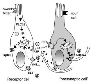
What did the article by Heath et al. (2006) actually show? Well, 2 hours after non-depressed people were given paroxetine (Paxil), an SSRI (selective serotonin reuptake inhibitor), or reboxetine (not approved for use in the US or Canada), a NARI (noradrenegic reuptake inhibitor), they were given a taste test. More specifically,
Subjects were informed of which taste modality (sweet, bitter, salt, or sour) they were receiving but were given no indication of whether the tester thought they would be able to recognize the taste or not. Low concentrations of one tastant may be misidentified as another; to avoid this potential confounding effect, subjects were informed as to the taste stimulus. ...Tastants were applied in solution to the tip of the tongue, using a cotton bud saturated with the solution at room temperature, and placed on the tongue for ~5 sec. Without closing their mouths, the subjects were asked to indicate whether or not they could taste the stimulus at that concentration.
The 20 participants visited the lab 3 times and were tested with varying concentrations of sucrose, quinine, NaCl, and HCl before and then 2 hrs after receiving either paroxetine, reboxetine, or placebo. Ultimately, what happened is that the SSRI increased sensitivity to both sweet and bitter tastes, while the NARI increased sensitivity to bitter and sour (see details below).
Heath TP, Melichar JK, Nutt DJ, Donaldson LF (2006). Human taste thresholds are modulated by serotonin and noradrenaline. J. Neurosci. 26: 12664-12671.

Circumstances in which serotonin (5-HT) and noradrenaline (NA) are altered, such as in anxiety or depression, are associated with taste disturbances, indicating the importance of these transmitters in the determination of taste thresholds in health and disease. In this study, we show for the first time that human taste thresholds are plastic and are lowered by modulation of systemic monoamines. Measurement of taste function in healthy humans before and after a 5-HT reuptake inhibitor, NA reuptake inhibitor, or placebo showed that enhancing 5-HT significantly reduced the sucrose taste threshold by 27% and the quinine taste threshold by 53%. In contrast, enhancing NA significantly reduced bitter taste threshold by 39% and sour threshold by 22%. ...
Thus, the taste system turned out to be more plastic than expected. The authors suggest that 5-HT and NA levels play an important role in setting taste thresholds. They present an elaborate model (with cool cartoon taste receptors, below) of how these effects might be mediated at a cellular level (in the taste buds, not in the brain).


But antidepressants typically take 4-6 weeks to work, not 2 hrs. And what does all this have to do with drug screening in depressed people, anyway? That's the more controversial part, since at the outset they mention the "monoamine theory of depression" which we all know to be a simplistic idea [but see the recent article by Meyer et al. 2006, which explains everything in terms of elevated levels of MAO-A (monoamine oxidase A), a catabolic enzyme.]

Nonetheless, Heath et al. bravely contend (in the press materials) that their results do have direct relevance in developing a "taste taste" for prescribing the correct medications. In the journal article, however, they only acknowledge that taste disturbances are often noted in depression.

The second news item is based on documents released by the FDA:
Antidepressants boost young adults' suicidal risks

WASHINGTON (AP) -- Using antidepressants increases the risk of suicidal thoughts and behavior among young adults but lessens it for seniors, the Food and Drug Administration says.

The effects of antidepressants on adults from 25 to 64 were mixed, so much so that the FDA would only conclude the drug had a neutral effect on suicidal behavior for them but possibly lowers the risk of suicidal thoughts.

The information came from a mass review of 372 studies involving roughly 100,000 patients and 11 drugs, including Lexapro, Zoloft, Prozac and Paxil. The FDA analysis will be incorporated in future changes to antidepressant labels, but the agency wants to first discuss its plans with outside advisers.

In 2004, the FDA ordered strong warnings about the pediatric risk of suicidal tendencies put on antidepressant labels, and began analyzing whether adults face a similar risk, in part by requesting data from drug companies.

It now appears there is an increased risk among adults between the ages of 18 and 25.

. . .
I have nothing funny to say about that.

Subscribe to Post Comments [Atom]

I Should've Forgotten That


The illustration above is from a brand new paper by Feredoes, Tononi, and Postle that used the latest in transcranial magnetic stimulation (TMS) technology to transiently disrupt bits of cortex (targeted regions indicated by markers) during a verbal working memory task. They were particularly interested in a phenomenon called proactive interference, in which previously remembered information infereres with the ability to remember new material. Overcoming PI involves the inhibition of recently remembered but currently irrelevant information.

Now back to TMS. The authors used repetitive TMS (rTMS) to test whether the left inferior frontal gyrus (IFG) was necessary for overcoming proactive interference. [They had their reasons for suspecting this, based on the existing literature.] They also applied rTMS to brain regions that were not expected to affect proactive interference (postcentral gyrus, motor cortex, and supplementary motor area) to control for non-specific effects.

Here's the coolest part (my emphasis):
rTMS was delivered with a Magstim Standard Rapid magnetic stimulator fit with a 70 mm figure 8 air-cooled stimulating coil (Magstim Co., Whitland, Wales, U.K.). Localization of the stimulating coil was accomplished via infrared-based frameless stereotaxy (eXimia Navigated Brain Stimulation, Nexstim, Helsinki, Finland). For each subject resting motor threshold (MT) was determined as the intensity at which single pulses applied over the hand area of right M1 produced a visible muscle twitch in five out of ten consecutive trials.
eXimia NBS Navigation System, as featured in Medgadget!

The results conformed to their prediction [but you might not believe it based on the figure below, particularly since non-rTMS performance in the left PCG condition was so poor]: rTMS over left IFG (but not other areas) impaired accuracy on working memory trials in which proactive interference was an issue (denoted by blue arrows), but not on other types of trials.


Feredoes E, Tononi G, Postle BR. (2006). Direct evidence for a prefrontal contribution to the control of proactive interference in verbal working memory. Proc. Natl. Acad. Sci. Published online before print December 6, 2006.

Controlling the effects of proactive interference (PI), the deleterious effect of prior mental activity on current memory representations, is believed to be a key function of the prefrontal cortex. This view is supported by neuroimaging evidence for a correlation between the longer reaction times caused by high PI conditions of a working memory task and increased activity in left inferior frontal gyrus (IFG) of the prefrontal cortex. An alternative that has never been ruled out, however, is that this left IFG effect may merely reflect sensitivity to such nonspecific factors as difficulty and/or time on task. To resolve this confound, we applied the interference methodology of repetitive transcranial magnetic stimulation (rTMS) to the left IFG and two control regions while subjects performed delayed letter recognition. rTMS was guided with high-resolution magnetic resonance images and was time-locked to the onset of the memory probe. The effect of rTMS, a disruption of accuracy restricted to high-PI probes, was specific to the left IFG. These results demonstrate that unpredictable, phasic disruption of the left IFG selectively disrupts control of responses to high-conflict verbal working memory probes, and they conclusively reject nonspecific alternative accounts.
And below is what the authors have to say about their study in the popular press:
Controlling confusion -- Researchers make insight into memory, forgetting

. . .

"Psychologists have known for decades that the intuitive notion of decay is probably less of a factor in forgetting than is interference," he says. Interference occurs, he says, when "other remembered information disrupts, competes with or confuses the information that you want to remember."

Interference is always present, Postle says, but we don't always notice it.

. . .

In the current study, volunteers read a group of letters ("F, B, P, X"), and were asked a few seconds later whether a particular letter had appeared in the most recent group (Did you just see a "Z"). In this type of test, having seen a "Z" in the string-before-last causes interference that makes the task more difficult. The subjects take longer to respond, and are more likely to incorrectly say "yes."

The research set-up was designed to be a simplified version of many everyday memory challenges, says Postle. Without a good sorting mechanism, our brains would be utterly confused by the vast amount of observations, ideas and memories that we have stored away. We might, for example, dial the phone number of the friend we just called rather than the one we intended to call.

In previous studies of interference, the IFG consistently lit up in brain scans, showing that it does something when the memory tries to deal with interference. But the IFG could simply be contributing some type of generic processing power to the task, says Postle.

However, the new study proved that the IFG is essential to blocking interference, he says, because accuracy plummeted when the IFG got a brief jolt of magnetic stimulation at the exact moment when the subject was confronting confusion.

Eventually, Postle hopes that locating the site of specific memory operations in the brain may help the millions of people with declining memories. "Understanding how the brain controls interference may be a first step to helping people with memory problems," he says.

The precise system used to target the magnetic pulse has many other applications in neuroscience research and treatment, Tononi adds. "TMS can be used not only to disrupt brain activity, but also to change it. If applied repeatedly, TMS can strengthen certain circuits that have become pathologically weak," he says.

Subscribe to Post Comments [Atom]

Monday, December 04, 2006

Encephalon - 12th Edition


A feast for the brain at AlphaPsy, with your host, Olivier.

Subscribe to Post Comments [Atom]

Sunday, December 03, 2006

Incredibly Obvious Headline of the Day

Testosterone May Affect Competitiveness

NO! Really?? I did not know this.

from APA online, Psychology in the News for the completely ignorant and uninformed:
United Press International - November 30, 2006

AUSTIN, Texas, Nov 30, 2006 (UPI via COMTEX) -- U.S. scientists have found a man's testosterone level might determine whether he's willing to get back into a competition after losing a challenge.

University of Texas at Austin psychology researchers Robert Josephs and Pranjal Mehta examined why some men back down after losing a contest, while others choose to challenge their opponent yet again. Their findings suggest the answer lies in what happens to a man's testosterone levels after the competition.

. . .

Among the men who lost the competition, 70 percent whose testosterone levels increased chose to compete again while 80 percent of those whose levels decreased declined the invitation.

I'm...I'm...rendered speechless by this fascinating new finding!

Subscribe to Post Comments [Atom]

LEARN TO TRUST YOUR OWN EYES

...or so says one of Jenny Holzer's truisms.

As an addendum to yesterday's post on Oxytocin and Mind Reading, I present to you the comments of Professor Paul Zak as a public service announcement, sponsored by The Neurocritic:

The synthetic oxytocin used in the experiments [by Kosfeld et al., 2005] has been around so long that it is available as a generic drug. It is no longer sold in the United States, though European women still use it to boost breastfeeding, said Paul Zak, one of the authors of the trust paper in Nature.

The dose needed to produce effects on trust was large -- subjects took about three teaspoonsful up their noses. But it appears to be quite safe, said Zak, who is director of the center for Neuroeconomic Studies at Claremont Graduate University in California.

The biggest side effect is that perhaps 20 percent of the men who take it get erections, he said, and, of course, pregnant women would want to avoid it because it could trigger contractions.

And in case you were wondering whether trust in a bottle would be worth your $49.95 for a 2 month supply, Zak had this to say:
On that score, a body spray on the market called ''Liquid Trust," is advertised as containing oxytocin that will induce unconscious trust in all who encounter you. But Zak said it's ''totally bogus," because sniffing oxytocin from someone's shirt collar will not get enough of the hormone to the brain. It's also available without a prescription -- unlike the real stuff -- he said, and overpriced: ''Liquid Trust" costs $49.95 for a two-month supply, while Zak and his colleagues made their inhalers for about $5 each.
The quotes above are from a great article in the Boston Globe online, Feeling shy, afraid of strangers? Hormone under study may help.

Remember: Don't Place Too Much Trust In Experts.

Subscribe to Post Comments [Atom]

Saturday, December 02, 2006

Oxytocin and Mind Reading...


...or, I Can See It In Your Eyes.

No, not that kind of "mind reading," but the ability to decode another person's mental state, also called theory of mind. In a study by Domes and colleagues (2006), the neurohormone oxytocin, peddled as the "trust drug" by many shady internet dealers, was administered to male subjects performing a theory of mind task.

[As an aside, nasally administered oxytocin does apparently cross the blood brain barrier designed to keep out foreign agents. I wasn't sure it crossed the BBB, but there you go.]

Anyway, the Reading the Mind in the Eyes task (see Baron Cohen... no, no joking! the OTHER Baron-Cohen) was administered after oxytocin or placebo. The RMET
tests a specific facet of mind-reading, that is, inference of the internal state from subtle affective facial expressions rather than mind-reading in general. Because the RMET was originally developed to measure severe impairments in mindreading capability in adults with autism spectrum disorders, we circumvented possible ceiling effects in healthy subjects by dividing the 36 items into two subsets of easy and difficult items.
Lo and behold, there was a tiny but statistically significant improvement in performance for oxytocin vs. placebo:
Compared with placebo, oxytocin improved performance on the RMET in 20 of the 30 participants. This resulted in a significant mean increase of approximately 3% correct responses (mean +/- SD: placebo: 69.4 +/- 8.1; oxytocin: 72.4 +/- 8.6; t=2.18, p=.019, one-sided).
But a breakdown of hard-to-read vs. easy-to-read eyes revealed that the benefit was entirely for the former.
Domes G, Heinrichs M, Michel A, Berger C, Herpertz SC. (2006). Oxytocin improves "Mind-Reading" in humans. Biol Psychiatry. Nov 28; [Epub ahead of print]

BACKGROUND: The ability to "read the mind" of other individuals, that is, to infer their mental state by interpreting subtle social cues, is indispensable in human social interaction. The neuropeptide oxytocin plays a central role in social approach behavior in nonhuman mammals. METHODS: In a double-blind, placebo-controlled, within-subject design, 30 healthy male volunteers were tested for their ability to infer the affective mental state of others using the Reading the Mind in the Eyes Test (RMET) after intranasal administration of 24 IU oxytocin. RESULTS: Oxytocin improved performance on the RMET compared with placebo. This effect was pronounced for difficult compared with easy items. CONCLUSIONS: Our data suggest that oxytocin improves the ability to infer the mental state of others from social cues of the eye region. Oxytocin might play a role in the pathogenesis of autism spectrum disorder, which is characterized by severe social impairment.
But...but...supposedly women will trust you more after spraying some oxytocin in their vicinity. Now we find out that they'll also be better able to read your mind.1

1 That's assuming the results from male participants will also hold for females.

Subscribe to Post Comments [Atom]

Friday, December 01, 2006

THC and Memory


Recently it was announced that tetrahydrocannabinol (THC), the active ingredient in cannabis, may help prevent Alzheimer's disease:

Eubanks LM, Rogers CJ, Beuscher IV AE, Koob GF, Olson AJ, Dickerson TJ, Kim D. Janda (2006). A molecular link between the active component of marijuana and Alzheimer's Disease pathology. Mol. Pharm. ASAP Web Release Date: 09-Aug-2006.

. . . Here, we demonstrate that the active component of marijuana, delta-9-tetrahydrocannabinol (THC), competitively inhibits the enzyme acetylcholinesterase (AChE) as well as prevents AChE-induced amyloid beta-peptide (A-beta) aggregation, the key pathological marker of Alzheimer's disease. Computational modeling of the THC-AChE interaction revealed that THC binds in the peripheral anionic site of AChE, the critical region involved in amyloidgenesis. Compared to currently approved drugs prescribed for the treatment of Alzheimer's disease, THC is a considerably superior inhibitor of A-beta aggregation, and this study provides a previously unrecognized molecular mechanism through which cannabinoid molecules may directly impact the progression of this debilitating disease.


see also:
Marijuana may block Alzheimer's
Tuesday, 22 February, 2005

Scientists showed a synthetic version of the compound may reduce inflammation associated with Alzheimer's and thus help to prevent mental decline.

. . .

Using cell cultures, the researchers confirmed that cannabinoids counteracted the activation of microglia and thus reduced inflammation.

[original research article by Ramirez et al., 2005]
However, even more recently, we've seen the report that THC is bad for your memory [what a surprise! but seriously, see Ranganathan & D'Souza, 2006 for a comprehensive review] because it disrupts synchronous cell firing in the hippocampus:
Robbe D, Montgomery SM, Thome A, Rueda-Orozco PE, McNaughton BL, Buzsaki G. (2006). Cannabinoids reveal importance of spike timing coordination in hippocampal function. Nat Neurosci. 9: 1526-1533.

Cannabinoids impair hippocampus-dependent memory in both humans and animals, but the network mechanisms responsible for this effect are unknown. Here we show that the cannabinoids Delta(9)-tetrahydrocannabinol and CP55940 decreased the power of theta, gamma and ripple oscillations in the hippocampus of head-restrained and freely moving rats. These effects were blocked by a CB1 antagonist. The decrease in theta power correlated with memory impairment in a hippocampus-dependent task. By simultaneously recording from large populations of single units, we found that CP55940 severely disrupted the temporal coordination of cell assemblies in short time windows (less than 100 ms) yet only marginally affected population firing rates of pyramidal cells and interneurons. The decreased power of local field potential oscillations correlated with reduced temporal synchrony but not with firing rate changes. We hypothesize that reduced spike timing coordination and the associated impairment of physiological oscillations are responsible for cannabinoid-induced memory deficits.
So it seems that short-term episodic and working memory (Ranganathan & D'Souza, 2006)...
cannabinoids impair all stages of memory including encoding, consolidation, and retrieval
...can be compromised with a long-term goal of preventing Alzheimer's disease!

More news stories!

Marijuana's Key Ingredient Might Fight Alzheimer's

Why Marijuana Impairs Memory

Marijuana's High Times Not Memorable with Neurons Out of Sync

References

Ramirez BG, Blazquez C, Gomez del Pulgar T, Guzman M, de Ceballos ML. (2005). Prevention of Alzheimer's disease pathology by cannabinoids: neuroprotection mediated by blockade of microglial activation. J Neurosci. 25: 1904-1913.

Ranganathan M, D'Souza DC. (2006). The acute effects of cannabinoids on memory in humans: a review. Psychopharmacology 188: 425-444.

Subscribe to Post Comments [Atom]

eXTReMe Tracker